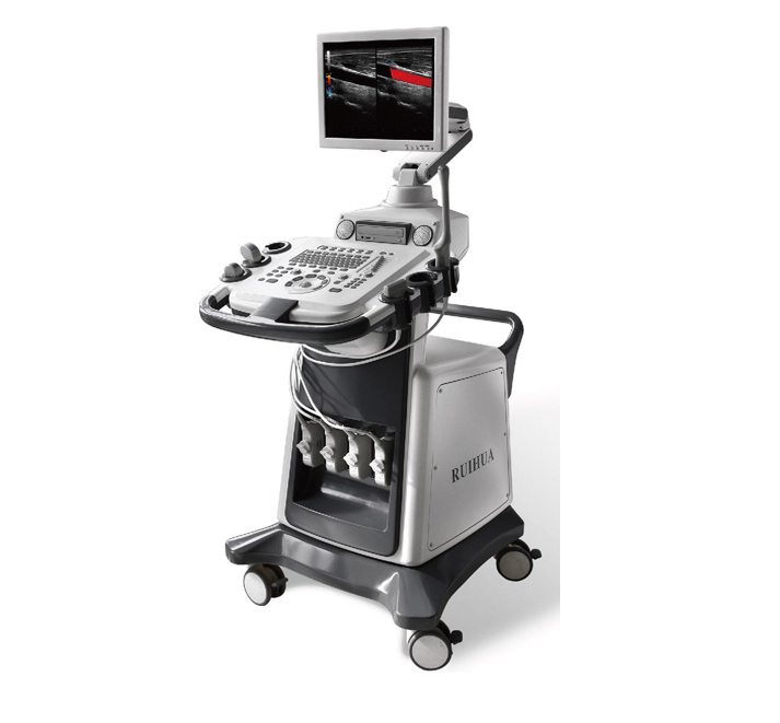
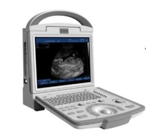
描述
Detailed introduction:
Scanning method
Electronic convex array, electronic linear array
Display mode: black and white images: B, 2B, 4B, left and right B/M, B/D, PW, M, B mode partial enlargement
Blood flow image CFM: B/C, BC/B, B/BC
Spectrum: B/D, B/C/D, PW
Color blood flow image adjustment parameters
Doppler frequency, sampling frame position and size, baseline, color gain, deflection angle, wall filtering, accumulation times, etc.
Probe type
Probe socket 4
Probe frequency: 2.0MHz ~ 10.0MHz, 8 levels of frequency conversion
Types of probes: large convex, slightly convex, linear array, cavity
Sound power: 16-level output sound power adjustable
Display depth: Using the continuous multi-stage adjustable encoder
Focusing: Electronic focusing + acoustic lens focusing
Launch focus: single focus, dual focus, triple focus and quad focus
Front-end reception uses continuous dynamic focusing
Grayscale 256 grayscale
Peripheral interface: DICOM interface (network interface), USB interface, RS232 interface
Optional: laser printer, video image printer
Obstetric measurement and calculation functions: fetal sac (GS), double top diameter (BPD), head and hip length (CRL), femur length (FL), humerus length (HL), transverse abdominal diameter (TAD), spine length (LV), Pillow frontal diameter (OFD), abdominal circumference (AC), head circumference (HC) estimate the length of the neck transparent layer, gestational age and expected date of delivery
Signal Processing and Doppler:
Dynamic filtering and quadrature demodulation
With total gain adjustment
Gain adjustment: 8-stage TGC
The total gains of type B, C and D are adjustable respectively
Black and white image gain and color flow gain are adjustable
Doppler stereo output volume adjustment 0-100%
Doppler baseline adjustment level 6
The pulse repetition frequency can be adjusted separately: CFM PWD
With D line speed adjustment


